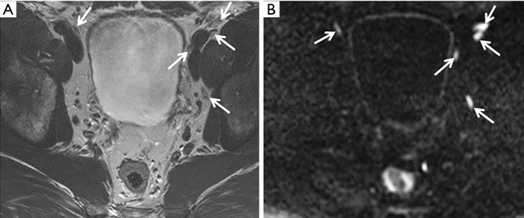
Figure 1.
A 68-year-old, PSA 6.3 ng/mL referred for MRI pre-biopsy. No primary tumour detected within the gland. Multiple small volume normal nodes are identified (arrows) on T2-weighted imaging (A), with the nodes being more easily appreciated on the b-1400 diffusion-weighted imaging (B). PSA, prostate-specific antigen; MRI, magnetic resonance imaging.

