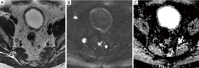
Figure 3.
A 71-year-old, PSA 51 ng/mL referred for MRI pre-biopsy. Large volume tumour with extra-capsular extension (not shown), Gleason 4+5 on targeted biopsy. Right and left internal iliac nodes involved, depicted on T2 and b-1400 imaging (A,B), with marked restricted diffusion on ADC maps, value 0.457×10⁻³ mm2/s (arrow) (C). PSA, prostate-specific antigen; MRI, magnetic resonance imaging; ADC, apparent diffusion coefficient.

