
Figure 2.
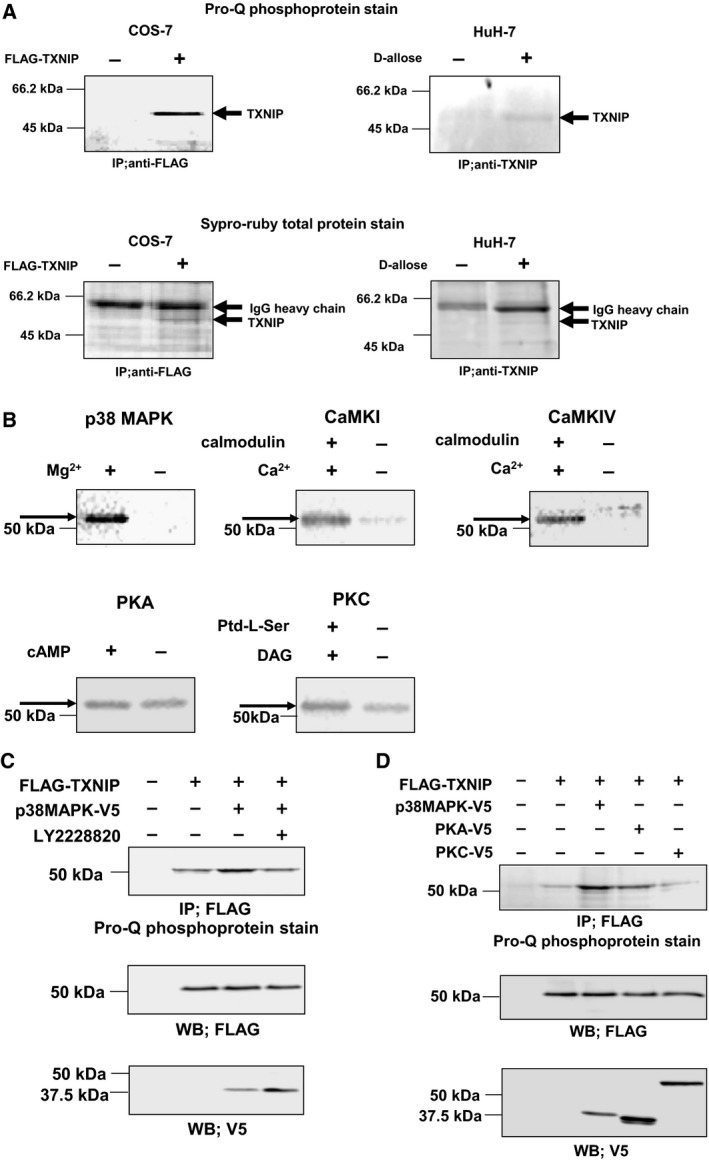
TXNIP is phosphorylated by p38 MAPK. (A) Phosphorylation of TXNIP in COS‐7 cells and HuH‐7 cells. COS‐7 cells were transfected with expression plasmid for FLAG–TXNIP, and cell lysate was immunoprecipitated with anti‐FLAG agarose gel. HuH‐7 cells were treated with 50 mm d‐allose for 48 h, and cell lysate was immunoprecipitated with anti‐TXNIP/protein G, and separated by SDS/PAGE. The phosphorylation of TXNIP was detected by Pro‐Q phosphoprotein gel stain. (B) In vitro phosphorylation analysis of TXNIP by autoradiography. Affinity‐purified FLAG–TXNIP protein was incubated in the presence of each kinase and γ‐[32P]ATP, and phosphorylated TXNIP was detected. (C) Phosphorylation of TXNIP by p38 MAPK in COS‐7 cells. COS‐7 cells were transfected with expression plasmid for FLAG–TXNIP and p38 MAPK–V5. Cells were pretreated with LY2228820 (500 nm; 2 h) as indicated. The phosphorylation of TXNIP was analyzed by immunoprecipitation followed by the Pro‐Q phosphoprotein gel stain. (D) Phosphorylation of TXNIP by kinases in COS‐7 cells. COS‐7 cells were cotransfected with expression plasmids for FLAG–TXNIP and each V5‐tagged kinase. The phosphorylation of TXNIP was analyzed by immunoprecipitation followed by Pro‐Q phosphoprotein gel stain.