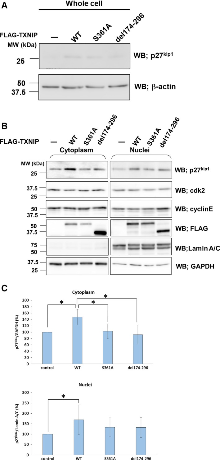
Figure 7.

Phosphorylation at Ser361 and C‐arrestin domain regulates the amount of p27kip1. HuH‐ 7 cells were transfected with TXNIP or its mutants in the expression plasmids. Whole‐cell lysates (A), cytoplasmic and nuclear fractions (B) of the transfected HuH‐7 cells were prepared for western blots. The antibodies used in Western blots are indicated in the figure. (C) Statistical analysis of western blots for p27kip1. Amount of p27kip1 in the transfected cells (TXNIP wild‐type or its mutants) was compared with that of non‐transfected cells for cytoplasm and nuclei. The relative amounts of p27kip1 are expressed as mean ± SD (*P < 0.05, t test, n = 4). GAPDH, glyceraldehyde 3‐phosphate dehydrogenase; WT, wild‐type.
