Figure 1.

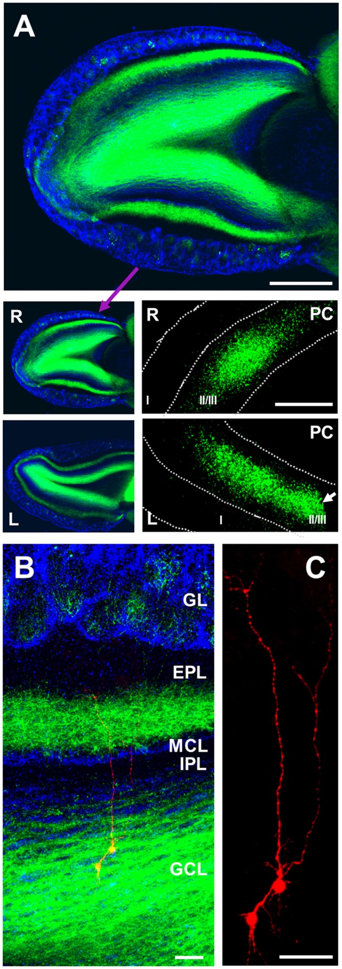
Anterograde labeling of cortico-bulbar glutamatergic axons. (A) Staining of channelrhodopsin-2 (ChR2; green) and DAPI (blue) showing distribution of labeled axons (green) in different layers of right (R) olfactory bulb (OB) with the densest labeling in granule cell layer (GCL), and second densest labeling in inner part of external plexiform layer (EPL). Limited fiber labeling is present in glomerular layer (GL), superficial EPL, internal plexiform layer (IPL) and mitral cell layer (MCL). Left (L) OB is also shown (low panel) a similar distribution of labeled axons from olfactory cortex (OCX). Note that left bulb was from cutting level of around 1,000–1,350 μm, and right bulb from around 300–650 μm to dorsal surface of OB. The right and left OCX with infected pyramidal cells are also shown (low panels), but the left one was cut to reduce slice size during the cutting process (white arrow). Scale bar: 400 μm in upper panel, and 1,000 μm in lower panel. (B) Triple staining of DAPI (blue), ChR2 (green) and biocytin (red) filling two patch clamped GCs with their somata located in the GCL and apical dendritic trunks projecting to ramify in the EPL. (C) Higher magnification showing the biocytin filled cells exhibit the classic dendritic arbor of GCs. Scale bar in (B,C): 50 μm.
