
Figure 2.
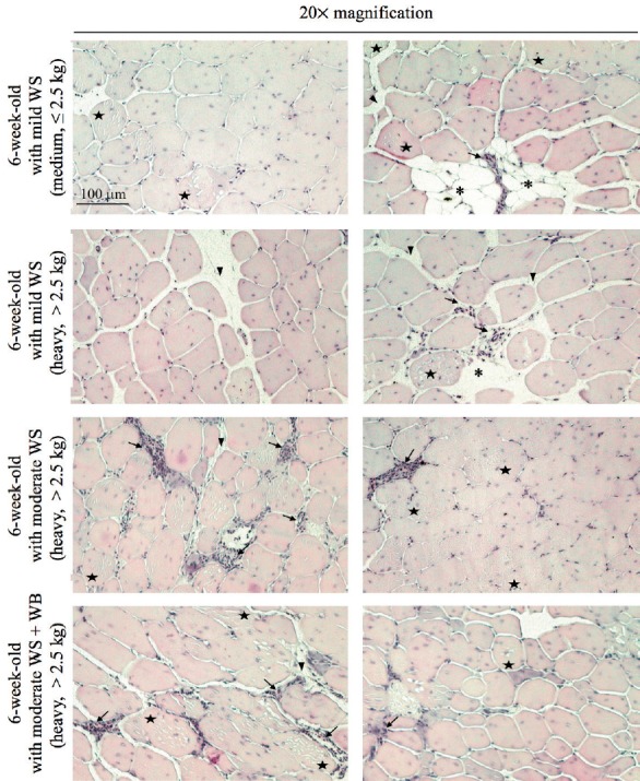
Histological images of the pectoralis major muscles depicted at 20× magnification. The breast muscle samples were collected from the broilers at the age of 6 weeks exhibiting various white striping (WS) severity with or without wooden breast (WB) condition. The broilers were graded as “medium” and “heavy” based on the carcass weight (medium, weight ≤2.5 kg; heavy, weight >2.5 kg). The fibers were eosin-stained and found as the polygonal structure. The nuclei were detected peripherally and internally in myofibers. Adipocytes (asterisks), macrophage deposition (arrows), and connective tissues (arrow heads) were found among the muscle fibers. Examples of necrotic fibers, observed as internal cell lesions, are marked with five-point stars. Scale bar = 100 μm for all images.