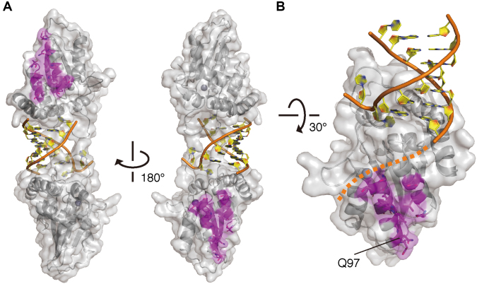
Figure 6.
A view of the Vif-interaction interface in the cpzA3H dimer. (A) Two separate A3H sites are responsible for Vif binding. cpzA3H and dsRNA are represented in gray ribbons with a transparent surface and orange/yellow ribbons with nucleobases, respectively. The residues (magenta sticks) critical for HIV-1–Vif interaction are mapped on the dimer structure. (B) Highlighted view of the Vif-binding interface and its unique residue Q97 in the chimpanzee-bonobo lineage. Residue Q97 is one of the most important determinants for the differential sensitivity of HIV-1/SIVcpz Vifs. A putative path for the extended ssRNA portion is also shown (orange dashed line).

