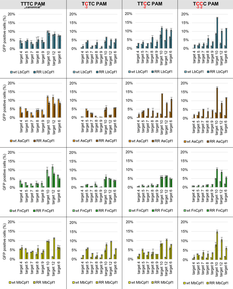
Figure 11.
Activity of WT and RR mutants on targets with TYYC PAM sequences in N2a cells. We compared the activity of WT and RR mutant Cpf1 nucleases on targets with TYYC PAM sequences (first column: TTTC, second column: TCTC, third column: TTCC, fourth column: TCCC) in a GFxFP assay. Percentages of GFP positive cells counted above the background level resulting from the action of As- (orange), Lb- (blue), Fn- (green) and MbCpf1 (yellow) are shown. The target vectors along with the corresponding nuclease vectors were transfected into N2a cells and GFP positive cells were counted 2 days post-transfection. All samples were also cotransfected with an mCherry expression vector to monitor the transfection efficiency and the GFP signal was analyzed within the mCherry positive population. The background fluorescence was estimated by using a crRNA-less, inactive LbCpf1 nuclease expression vector as negative control and was subtracted from each sample. Three parallel transfections were made for each case. Error bars show the mean ± standard deviation of percentages measured in n = 3 independent transfections. See also Supplementary Figures S17–S21.

