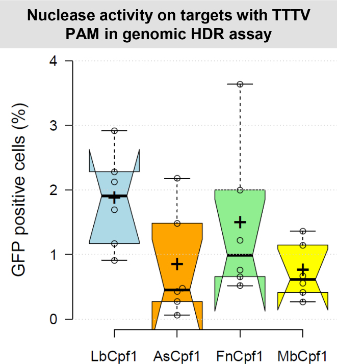
Figure 2.
Cpf1 nucleases induced HDR at various genomic cleavage sites in N2a cells. Percentages of GFP fluorescent cells after HDR mediated integration of a donor GFP cassette are shown. The efficiencies of Cpf1 nucleases (blue – LbCpf1, orange – AsCpf1, green – FnCpf1, yellow – MbCpf1) in inducing HDR mediated integration were measured on six mouse doppel genomic targets. The nuclease vector and the homologous recombination donor molecule with the corresponding homologous arms were co-transfected into N2a cells. As negative control, the homologous recombination donor without the corresponding homologous arms was used. On the 14th day after transfection, GFP positive cells were counted. The corresponding negative control was subtracted from each sample. Three parallel transfections were made for each sample. Two days after transfection all the samples made on the same day showed similar GFP positive cell counts. This GFP fluorescence was used to normalize the results for variation in transfection efficiency. Tukey-type notched boxplots by BoxPlotR (53): center lines show the medians; box limits indicate the 25th and 75th percentiles; whiskers extend 1.5 times the interquartile range from the 25th and 75th percentiles; notches indicate the 95% confidence intervals for medians; crosses represent sample means; data points are plotted as open circles that correspond to the different targets tested. See also Supplementary Figure S6.

