Figure 3.
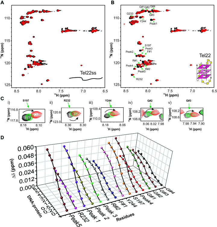
Interaction of RGG-box with the single stranded and G-quadruplex DNA monitored through NMR spectroscopy. (A) 2D 15N–1H HSQC spectrum of the free RGG-box (black) and in complex with Tel22ss at 1:6 protein to DNA molar ratio (red). No significant chemical shift perturbations were observed for this interaction. Single stranded Tel22ss is shown as a cartoon. (B) 2D 15N–1H HSQC spectrum of the RGG-box (black) and in complex with Tel22 at 1:6 protein to DNA molar ratio (red). Specific chemical shift perturbations were observed for several residues (marked with green arrows). A representative cartoon of monomeric G-quadruplex form of Tel22 is shown (only one conformation in K+ ion is shown). (C) A subset of residues of RGG-box that show specific chemical shift perturbation upon addition of Tel22 is shown. The RGG-box and Tel22 complex was in fast exchange (weak binding) as we observed continuous movement of resonance peaks upon addition of increasing amount of the Tel22 DNA. Three steps of titration at different protein to DNA ratios are shown: black at 1:0, green at 1: 1, and red at 1:6. (D) The titration curves showing chemical shift change plotted as a function of increasing DNA:protein ratio for 14 interacting residues of the RGG-box is shown.

