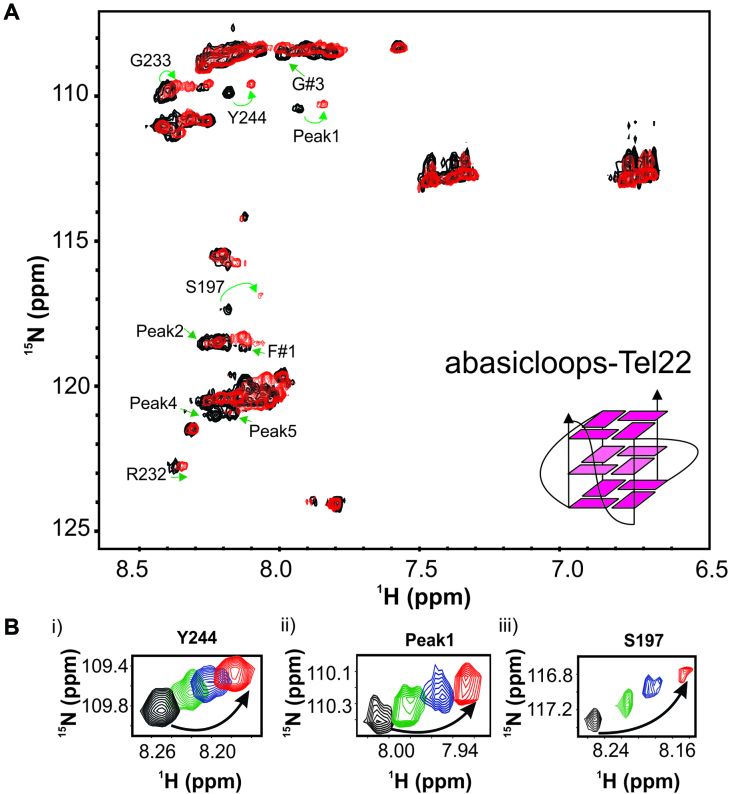
Figure 5.
The RGG-box interacts with the G-quadruplex with abasic deoxyribose loops. (A) 2D 15N–1H HSQC spectrum of the free RGG-box (black) and in complex with abasicloops-Tel22 DNA at 1:1 protein to DNA molar ratio (red). A representative cartoon of monomeric G-quadruplex form of abasicloops-Tel22 is shown. Specific chemical shift perturbations were observed for several residues (marked with green arrows). (B) A subset of residues of the RGG-box that show specific chemical shift perturbation upon addition of abasicloops-Tel22 is shown. The RGG-box and abasicloops-Tel22 complex was in fast exchange. Four steps of titrations at different protein to DNA ratios are shown: black at 1:0, green at 1:0.2, blue at 1:0.5 and red at 1:1.

