Figure 8.
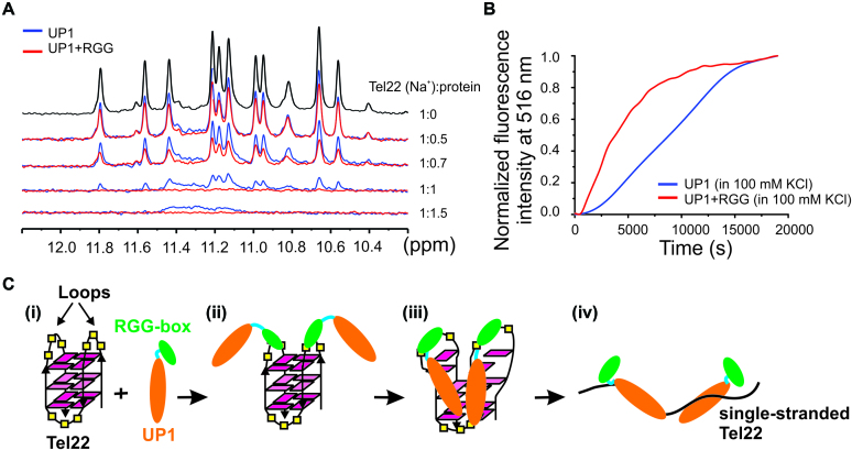
Telomere DNA G-quadruplex unfolding by UP1+RGG and UP1 monitored using NMR and fluorescence spectroscopy. (A) 1D 1H NMR spectra of Na+ form of Tel22 showing gradual loss of imino proton peaks upon titration with increasing concentrations of UP1 (blue) and UP1+RGG (red). (B) Unfolding of the 5′-FAM and 3′-TAMRA labeled K+ form of Tel22 DNA G-quadruplex (5′FAM-Tel22-TAMRA3′) by UP1 (blue) and UP1+RGG (red) monitored by observing the emission of FAM at 516 nM. 5′FAM-Tel22-TAMRA3′ DNA was mixed with 4 molar equivalents of UP1 or UP1+RGG and the emission spectrum was recorded over a time period. (C) Proposed model for RGG-box assisted recognition and unfolding of telomere DNA G-quadruplex unfolding by UP1+RGG.

