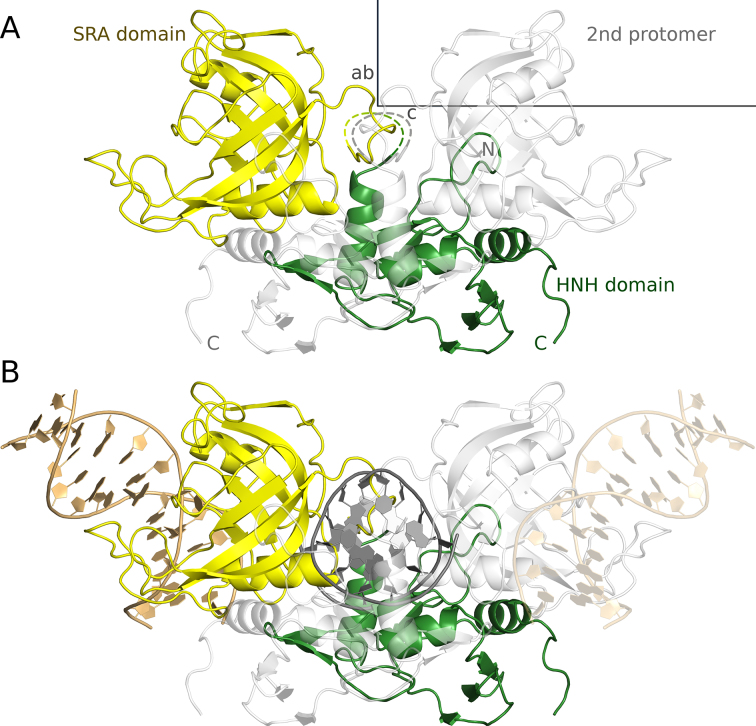
Figure 3.
Experimentally determined structure of TagI in the absence of DNA (A) and a model of TagI with separate DNA fragments bound to the SRA and HNH domains (B). The TagI protomer in the asymmetric unit is shown in yellow (SRA domain) and green (HNH domain), the crystallographic symmetry mate that completes the TagI dimer is shown in light gray. The fragment of the structure that is disordered in the crystal is indicated by a dashed line. The unit cell and the directions of the crystallographic axes are shown in gray. The DNA molecules that have been modelled in complex with the SRA domains of the dimer are shown in gold and light gold color, and the single modelled DNA molecule that is bound to the HNH dimer is shown in dark grey color.

