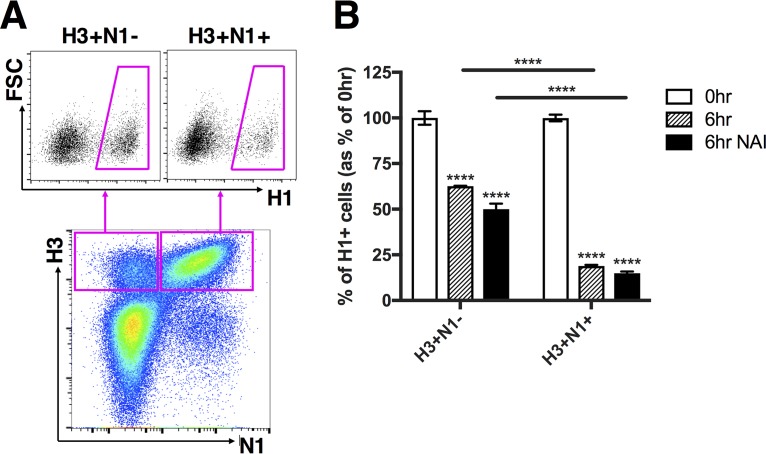
FIG 2.
Superinfection exclusion is more potent in infected cells that express NA but is independent of NA enzymatic activity. MDCK cells were infected with rH3N1 virus and were simultaneously (0hr) or sequentially (6hr) infected with rH1N2 virus; all infections were performed at MOI = <0.3 TCID50/cell. During the 5-h gap and 1-h adsorption of the secondary infection (rH1N2), cells were incubated in either medium alone or media with 1 µM zanamivir (NAI). (A) Representative FACS plots comparing H1+ frequencies between H3+ N1− and H3+ N1+ cells. (B) H1+ frequencies within H3+ N1− and H3+ N1+ cells following simultaneous (0hr) or sequential (6hr) infection. Values of both the 0-h and 6-h groups (with or without the presence of NAI) are normalized to the means of 0-h controls, and data are presented as mean values (n = 3 cell culture wells) ± standard deviations. ****, P < 0.0001 (t test).

