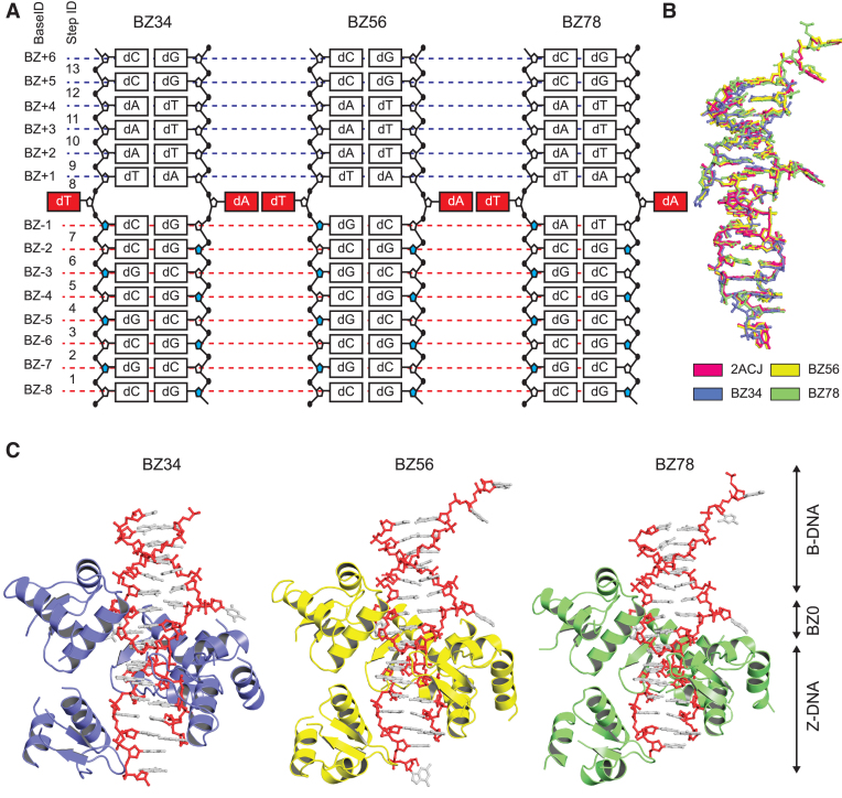
Figure 1.
Overall structures of BZ junctions. (A) Schematic diagram of DNA composition of BZ34, BZ56 and BZ78. The BZ junction positions are red and extruded from the stacking. The sugar backbones with syn conformations are colored in blue in the Z-DNA part. Base and step IDs are listed on the left side. (B) Structural comparison between the previously reported BZ junction structure (PDB code: 2ACJ) and three crystal structures of BZ junctions in this study. The structures of DNA are represented by ball and stick models. The structures of 2ACJ, BZ34, BZ56 and BZ78 are colored in pink, light blue, yellow and green, respectively. (C) The crystal structures of BZ34, BZ56 and BZ78. The hZαADAR1 domains of BZ34, BZ56 and BZ78 are colored in light blue, yellow and green, respectively. The DNA molecules of BZ34, BZ56 and BZ78 are represented by ball and stick models. The phosphate backbones of DNA are colored in red. The Z-DNA, BZ junction and B-DNA parts are labeled as Z-DNA, BZ0 and B-DNA using arrowed lines, respectively.

