Figure 6.
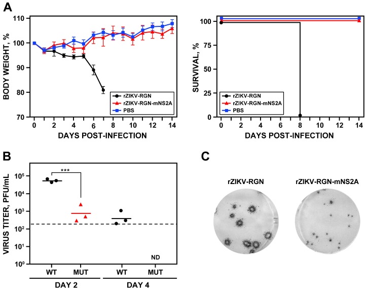
Pathogenesis of rZIKV-RGN-mNS2A in A129 mice. (A) Weight loss and mortality. Female 4-to-6-week-old A129 mice (five mice per group) were mock-infected (PBS) or infected s.c. in the footpad with 105 PFU of rZIKV-RGN or rZIKV-RGN-mNS2A, and body weight loss (expressed as the percentage of starting weight, left panel) and survival (right panel) were monitored daily during 14 days. Mice that lost more than 20% of their initial body weight or presented hind limb paralysis were humanely euthanized. Error bars represent standard deviations of the mean for each group of mice. (B) Viral titers in mice sera. Female 4-to-6-week-old A129 mice (six mice per group) were infected with 105 PFU of rZIKV-RGN (WT) or rZIKV-RGN-mNS2A (MUT) as described above, and viral titers in sera were determined at days two and four after infection (three animals per time point) by plaque assay and immunostaining using the pan-flavivirus E protein mAb 4G2. Symbols represent data from individual mice and bars the geometric means of viral titers. Asterisks indicate that the differences between rZIKV-RGN and rZIKV-RGN-mNS2A are statistically significant when data are compared using the unpaired t test (***, P < 0.001). ND: virus not detected. The detection limit of the assay (200 PFU/mL) is indicate as a dashed line. (C) Plaque phenotype. Vero cells at 90% confluence (6-well plate format) were infected with 25 PFU of rZIKV-RGN (left) or rZIKV-RGN-mNS2A (right) recovered from infected mice at day two post-infection and the plaque size evaluated by plaque assay and immunostaining using the pan-flavivirus E protein mAb 4G2.

