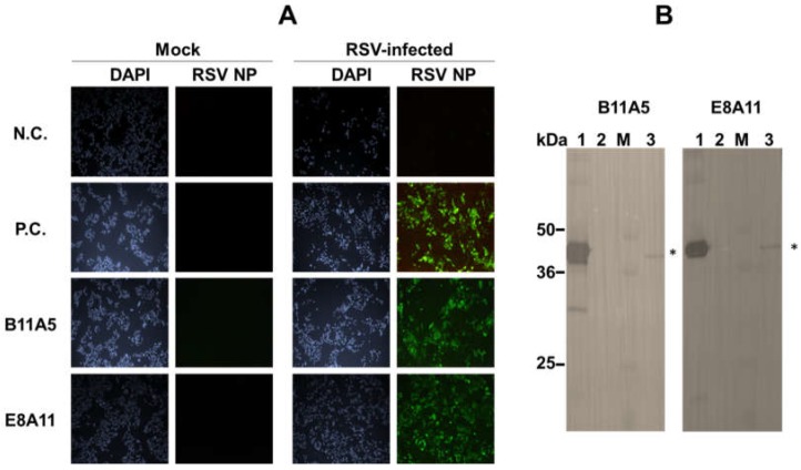
Figure 2.
Characterization of monoclonal antibodies against respiratory syncytial virus (RSV). (A) Cells were independently infected with the virus for 24 h and fixed with 4% paraformaldehyde. After fixation, cells were treated with lysis buffer and washed with phosphate buffered saline with 0.1% Tween-20 (PBST) three times. Green fluorescence was detected with a fluorescein isothiocyanate (FITC)-conjugated secondary antibody. N.C., negative sera; P.C., commercial anti-RSV NP antibody; Mock, uninfected. All images were acquired by resolution power setting with 100×. (B) Western blotting was conducted using an RSV-infected cell pellet. 1, purified RSV recombinant nucleoprotein (rNP; 5 µg/lane); 2, BSA (5 µg/lane); 3, marker; 4, RSV (1 × 10⁶ TCID50/mL)-infected cell pellet (4 µg/lane). Asterisk indicates RSV NP protein.

