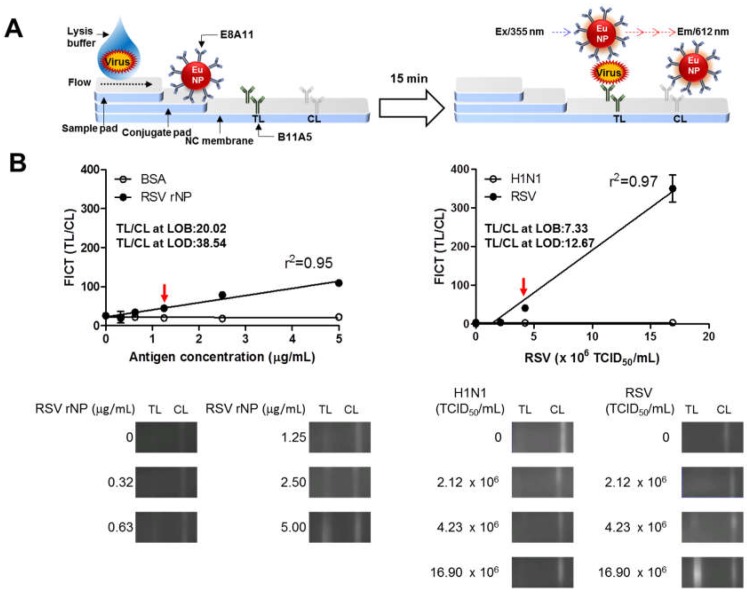
Figure 5.
Development of a rapid fluorescence diagnostic system for the detection of respiratory syncytial virus (RSV). (A) Schematic diagram of the rapid fluorescence diagnostic system employing a europium nanoparticle (Eu NP)-conjugated RSV-specific antibody. Fluorescence was measured for Eu NPs (excitation at 355 nm and emission at 612 nm). (B) Fluorescent immunochromatographic strip test (FICT) employing Eu NP-conjugated antibodies was tested for its limit of detection (LOD) against RSV rNP and RSV. The data (n = 3) are shown as the mean ± SD. Linear regression is shown with the line. The red arrow indicates the antigen concentration or virus titer at the LOD. Raw fluorescence images from the test line (TL) and control line (CL) of the FICT are shown in the bottom panel. The signals at the TL and CL were read with a portable strip reader, and the fluorescent values of TL/CL were computed and plotted on the graph.

