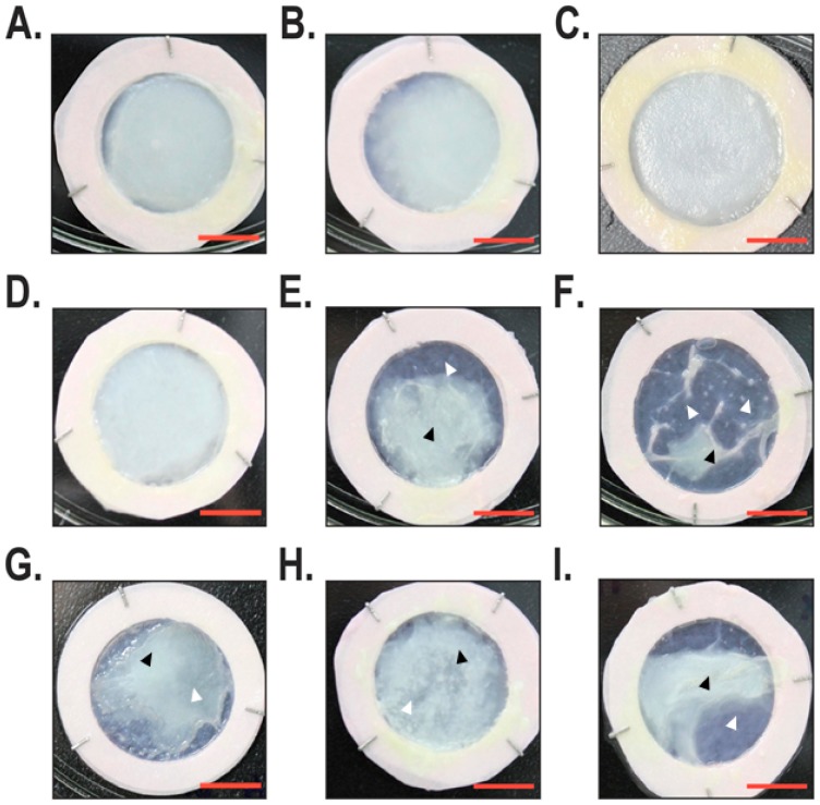
Figure 1.
Macroscopic analysis of the reconstructed skin substitutes. For each group, tissue-engineered skin substitutes were produced with three different cell populations. (A–C) Tissue-engineered skin substitutes produced with healthy fibroblasts and keratinocytes. (D–I) Tissue-engineered skin substitutes produced with fibroblasts and keratinocytes isolated from either non-lesional (D–F) or lesional (G–I) psoriatic skin. Scale bar = 1 cm. Black arrowheads indicate the position of protuberant regions, whereas white arrowheads position thinner regions of the reconstructed skin substitutes.

