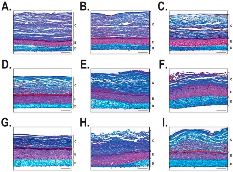
Figure 2.
Histological analysis of the reconstructed skin substitutes. Mason’s trichrome staining after 21 days of culture at the air-liquid interface. For each group, tissue-engineered skin substitutes were produced with three different cell populations. (A–C) Healthy skin substitutes. (D–F) Non-lesional psoriatic skin substitutes. (G–I) Lesional psoriatic skin substitutes. Scale bar = 100 µm. C: Stratum corneum; E: Epidermis; D: Dermis.

