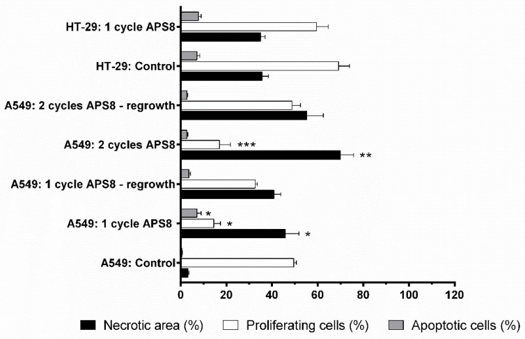
Figure 7.
Determination of tumor necrotic area and percentage of apoptotic and proliferating cells after APS8 treatment. Quantification was done from histological sections of human A549 xenograft lung adenocarcinoma and HT29 colon adenocarcinoma tumors after intratumoral treatment with APS8, as one or two cycles of treatment. Data are means ± SEM (n = 3). Necrotic areas evaluated on whole tumor sections. Percentages of apoptotic cells or proliferative cells were estimated in five fields in viable tumor areas. * P < 0.05, for A549 tumors untreated versus after one cycle of APS8 treatment; ** P < 0.05, for A549 tumors that responded to APS8 treatment versus those that started to regrow; *** P < 0.05, for A549 tumors treated with one versus two cycles of APS8 treatment.

