Figure 3.
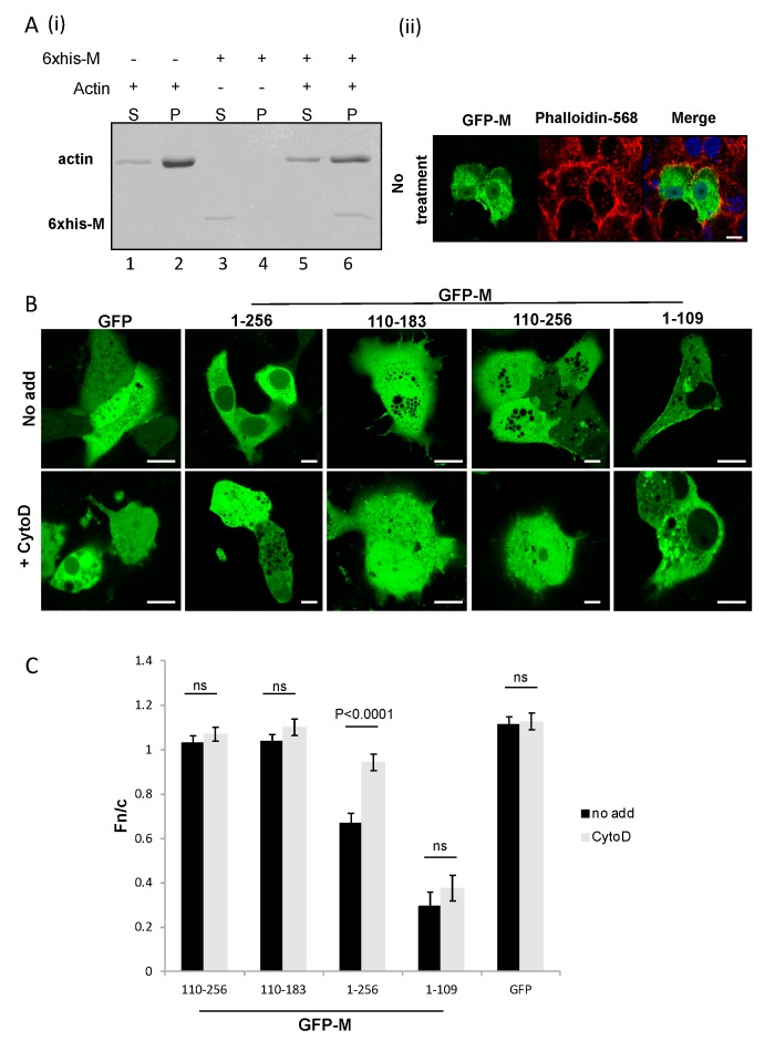
M binds polymerized actin. A(i) Recombinant purified M protein (6× His-M) was incubated with in vitro polymerized and monomeric actin, as described in the text. Samples were centrifuged to separate polymerized and monomeric actin. Supernatant (S) and pellet (P) fractions were analyzed with SDS-PAGE and visualized by Coomassie Brilliant Blue staining. The bands representing actin and RSV M are indicated. A(ii) A549 cells were cultured overnight and transfected to express full-length GFP-M (1–256). Transfected cells were left untreated and probed for visualization of the microfilament network (phalloidin-594). The colocalization of M and actin microfilaments is indicated in yellow (image labelled merge). Scale bars = 10 μm. (B) Cos-7 cells were cultured overnight and transfected to express GFP, GFP-M (1–256), or deletion mutants GFP-M (110–183) and GFP-M (110–256). Transfected cells were left untreated (no add) or treated with cytochalasin D (+CytoD) and imaged live using confocal microscopy. Representative images are shown. Scale bars = 10 μm. (C) Images such as those shown in (B) were analyzed by ImageJ to Fn/c. Data shown are mean ± SEM, n ≥ 15. Statistically significant differences between groups are shown, ns = non-significant.

