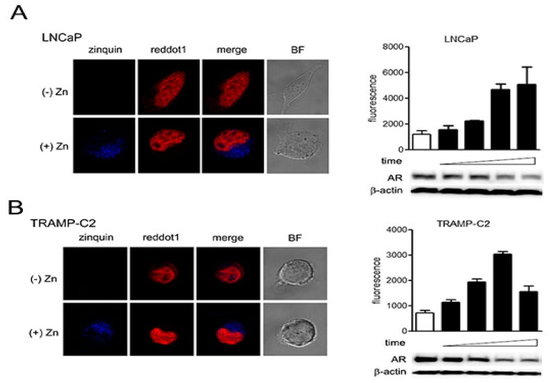
Figure 4.
Exogenous zinc is primarily localized to the cytoplasm. (A,B) (left panels) Cells were seeded in 4-well chambers to reach 60% confluence, then treated with zinc at 150 µM (LNCaP) or 75 µM (TRAMP-C2) for 6 h. Cells were then treated with 20 µM zinquin and incubated for 30 min at 37 °C. Cells were washed and treated with RedDot1 and observed by confocal microscopy. (A,B) (right panels) Fluorescence intensities were measured at 0, 1, 2, 4, and 6 h and are expressed as mean ± SD. Cells monitored by confocal microscopy were also used for protein analysis.

