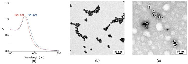
Figure 3.
Characterization of MIMO@LA@AuNPs. (a) Visible spectra of citrate-capped AuNPs (red line) and of the bioconjugate MIMO@LA@AuNPs (blue line). (b) Transmission Electron Microscopy (TEM) image of citrate-capped AuNPs. (c) TEM image of the bioconjugate MIMO@LA@AuNPs. The staining with 1% uranyl acetate allowed highlighting the protein shell as a white halo around each AuNP.

