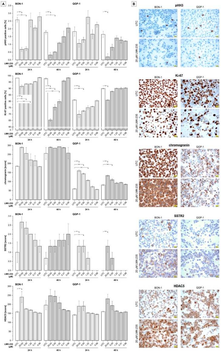
Figure 5.
Immunocytochemistry on cell blocks of LMK-235-treated pNET cells. (A) Semiquantitative evaluation of immunostaining for phosphohistone H3 (pHH3) and Ki-67 (% positive cells), chromogranin (score, based on multiplication of intensity and extensity; 0–300), SSTR2 (score; 0–3), and histone deacetylase-5 (HDAC5 score; 0–300). Cells were treated with 0.08–20 µM LMK-235 for 24 or 48 h and results represent mean values ± SEM of three independent images. Asterisks indicate p-values of <0.05 (one) or <0.01 (two) for comparison of treatments versus the corresponding untreated control (statistical results for group comparisons within treatments are not shown). (B). Representative images of the immunostainings for UTC and LMK-235-treated (20 µM, 24 h) samples. Magnification 400× for all images, scale bars indicates 20 µm. Abbreviations: UTC = untreated control.

