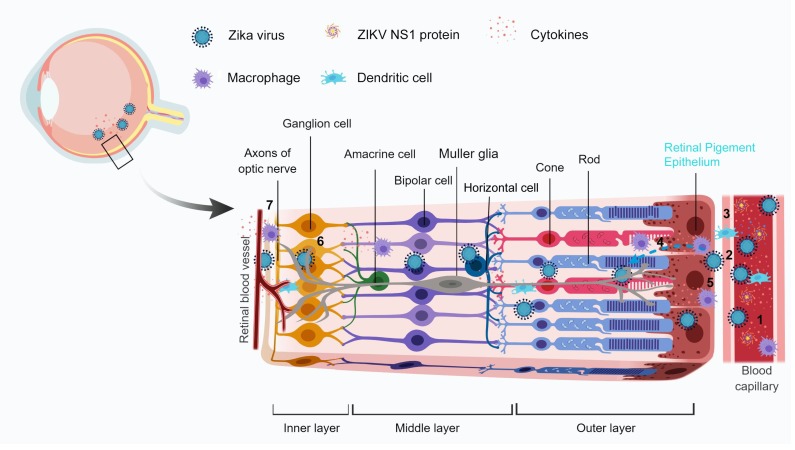
Figure 3.
Probable mechanisms for the breach of blood-retinal barriers by ZIKV. Upon infection and peak viremia, there is an increased circulation of Zika virus, ZIKV NS1 protein, and immune cells in the blood (1). The virus in the retinal blood capillaries infect the endothelial lining (2) and the immune cells reach the site of infection/inflammation by diapedesis through the capillaries (3). It is followed by infection of RPE, the cell lining the outer BRB, resulting in chorioretinal atrophy. The viral infections might cause a BRB weakening by decreasing intercellular junction integrity. Being a neurotrophic virus, at later stages ZIKV can infect retinal Muller glia or neurons inside of the eye (4). The complications are worsened with the involvement of immune cells which get activated upon infection and release a “cytokine storm” as an antiviral response which damages the host cells by altering the barrier integrity (5). The virus along with the circulating immune cells can cross the inner BRB (retinal blood vessels) and infect neuronal cells such as ganglion cells (6, 7). The Image has been created with BioRender software.

