Figure 1.
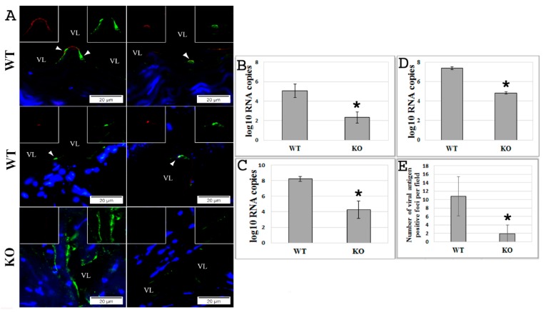
Absence of the exchange protein directly activated by cAMP 1 (EPAC1) gene attenuates Ebola virus (EBOV) infection ex vivo and in vitro. (A) Representative dual-target immunofluorescence (IF) staining of EBOV (red) and endothelial cell (EC)-specific von Willebrand factor (vWF; green) in sections of ex vivo aortic ring vasculatures prepared from Epac1-null (KO) and wild-type (WT) mice 72 h postinfection (p.i.) with EBOV. Nuclei of mouse cells were counterstained with DAPI (blue). Inserts depict split signals of EBOV (red) and vWF (green) from the colocalized areas (arrow heads). Scale bars, 20 µm. VL, vascular lumen. (B) The number of viral RNA copies detected in the media of ex vivo aortic ring vasculatures of KO and WT mice at 72 h p.i. with EBOV. N = 3 for each group. (C,D) The number of viral RNA copies detected in brain microvascular endothelial cells (BMECs) (C) and media (D) at 72 h p.i. with EBOV at an MOI of 0.5. N = 3 for each group. (E) The number of viral antigen-positive foci measured using IF microscopy in the monolayers of BMECs, which were isolated from KO and WT mice, at 72 h p.i. with EBOV at an MOI of 0.5. N = 30 for each group. * p < 0.005 compared with WT groups.

