Figure 2.
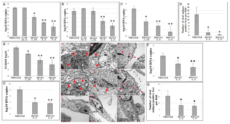
Pharmacological inactivation of EPAC1 protects human umbilical vein endothelial cells (HUVECs) from EBOV infection. (A,B) The number of viral RNA copies detected in Vehicle- (N = 5), ESI09- (N = 5), and H89-pretreated HUVECs (N = 3) (A) and media (B) at 72 h p.i. with EBOV at an MOI of 0.5. * p < 0.05, ** p < 0.005 compared with Vehicle groups. (C) The number of viral RNA copies detected in the media from Vehicle- (N = 3) and NY0123-pretreated (N = 3) HUVECs at 72 h p.i. with EBOV at an MOI of 0.5. N = 3 for each group. * p < 0.05, ** p < 0.005 compared with the Vehicle group. (D) The number of viral antigen-positive foci measured using IF microscopy in the monolayers of HUVECs at 72 h p.i. with EBOV at an MOI of 0.5. N = 30 for each group. * p < 0.05 compared with the Vehicle group. (E) Quantities of viral particles in media measured using the TCID50 assay, N = 3 for each group. * p < 0.05, ** p < 0.005 compared with the Vehicle group. (F–N) Representative electron microscopy (EM) detection of EBOV particles (arrow heads) in HUVECs pretreated with DMSO (F–K), ESI09 (L–M), and NY0123 (N) at 72 h p.i. with EBOV at an MOI of 0.5. EM images of 10 cells for each group were reviewed under EM. Scale bars, (F–K), 500 nm; (L–N), 2 µM. (O) and (P) HUVECs infected with EBOV at an MOI of 0.5 and treated with ESI09 and NY0123 at 5 µM 24 h later. The number of viral RNA copies detected at 72 h p.i. in the cells (O) and media (P). N = 3 for each group. * p < 0.05, ** p < 0.01 compared with Vehicle groups. (Q) The number of viral antigen-positive foci measured at 72 h p.i. using IF microscopy in the monolayers of HUVECs treated with ESI09 or NY0123 at 24 h p.i. N = 30 for each group. * p < 0.05 compared with the Vehicle group.

