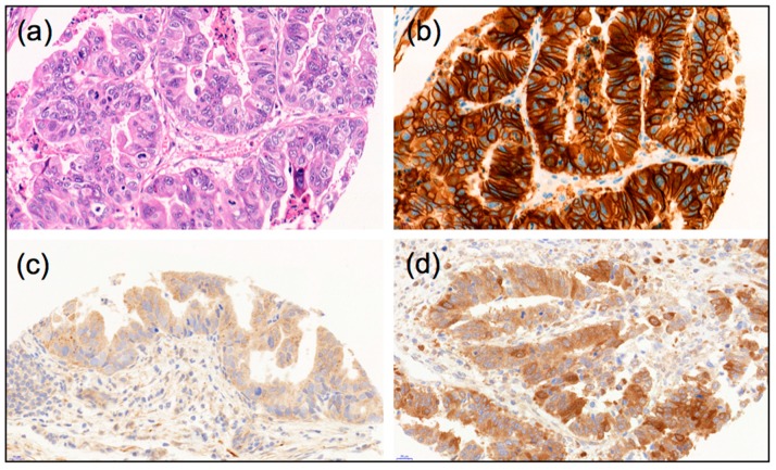
Figure 4.
Examples of morphology, autophagy markers and Her2 immunohistological stainings of esophageal adenocarcinomas: (a) Hematoxylin-Eosin stain; (b) Example of strong Her2-positive staining; (c) Example of LC3B high dot-like staining; (d) Example of p62 high cytoplasmic/dot like staining, (40× magnification each).

