Figure 1.
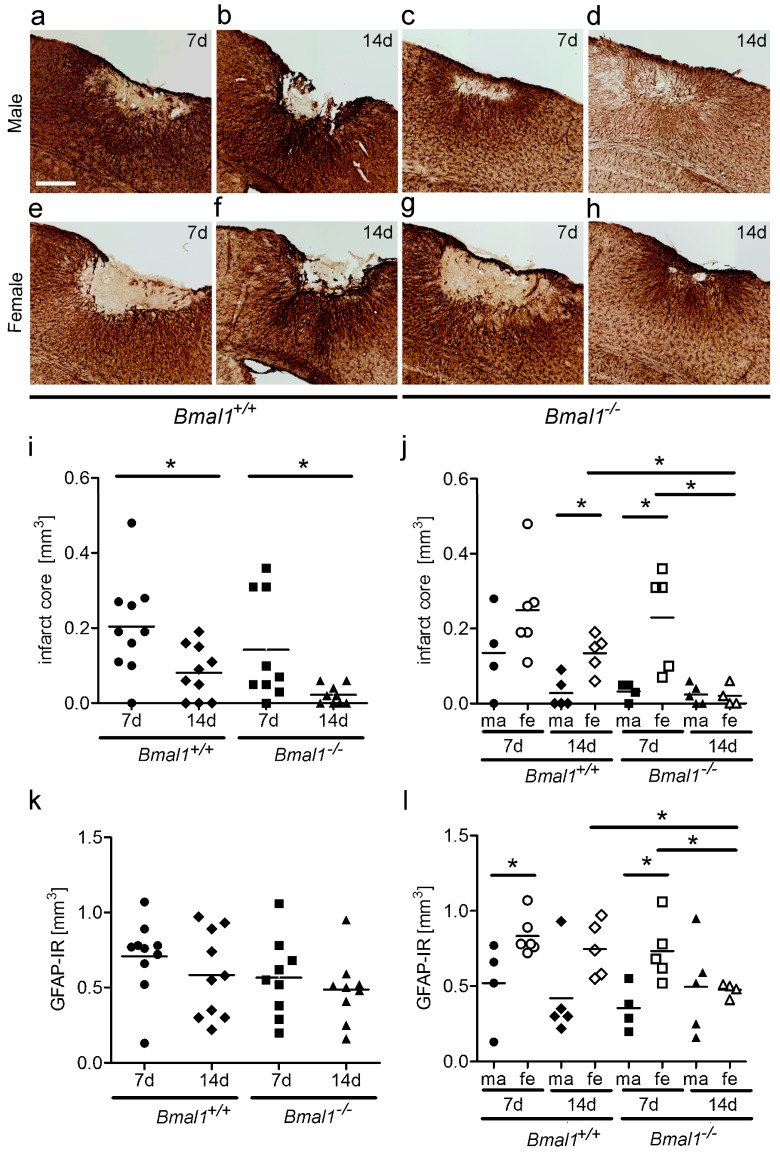
Effect of Bmal1-deficiency and sex on infarct core and astrocytes in the peri-infarct area. Infarct core and reactive astrogliosis in the peri-infarct area after focal cortical ischemia induced by photothrombosis (PT) were determined by GFAP-immunoreaction (IR). (a) Bmal1+/+ male 7 days (d) after PT; (b) Bmal1+/+ male 14 days after PT; (c) Bmal1−/− male 7 days after PT; (d) Bmal1−/− male 14 days after PT; (e) Bmal1+/+ female 7 days after PT; (f) Bmal1+/+ female 14 days after PT; (g) Bmal1−/− female 7 days after PT; (h) Bmal1−/− female 14 days after PT; (i) volume of infarct core in Bmal1+/+ and Bmal1−/− mice of both sexes (n = 9–10 per genotype); and (j) volume of infarct core in Bmal1+/+ and Bmal1−/− mice separated by sex (n = 4–5 per genotype and sex). (k) Volume of reactive astrogliosis in the peri-infarct area in Bmal1+/+ and Bmal1−/− mice of both sexes (n = 9–10 per genotype); (l) Volume of reactive astrogliosis in the peri-infarct area in Bmal1+/+ and Bmal1−/− mice separated by sex (n = 4–5 per genotype and sex). * p < 0.05, Mann Whitney-U Test. Scale bar = 300 µm.

