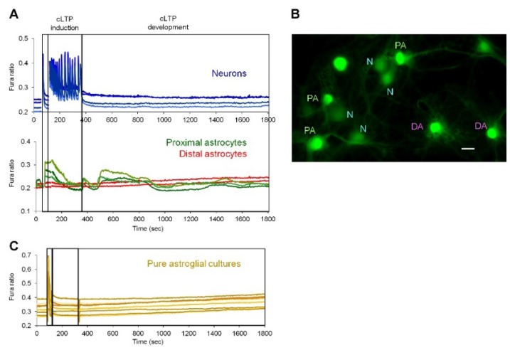
Figure 4.
Delayed Ca2+ signals in proximal-to-neurons astrocytes induced by cLTP. (A) Ca2+ signals were detected by Fura-2 single cells imaging and analyzed separately in neurons (blue traces) and astrocytes (green and red traces). Note appearance of delayed Ca2+ signals in astrocytes which are in close apposition with neurons (green traces in panel A and “PA” (Proximal Astrocytes) label in B) but not in distal astrocytes, which are not in contact with neurons (“N”) (red traces in panel A and “DA” label in panel B). Representative traces and image are shown of cLTP induction experiments from 2–3 coverslips (technical replicates) for each of three independent culture preparations (biological replicates). Bar, 10 μm. C, Ca2+ signals were not detected in pure astroglial cultures stimulated with cLTP protocol.

