Figure 4.
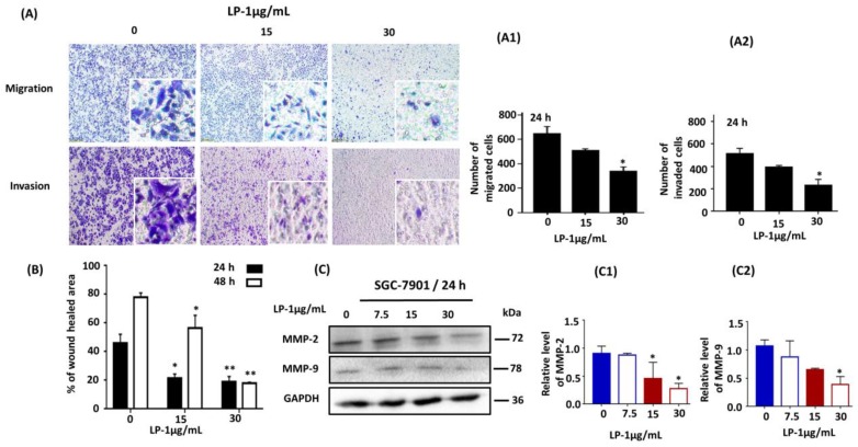
LP1 suppresses migration, invasion and wound healing in SGC-7901 cells. (A) Cell migration and invasion assays were performed with (0, 15, 30 µg/mL) of LP1 for 24 h. Images were captured by phase contrast microscope, scale bar 200 µm. (A1) A number of migrated cells quantified by using ImageJ software. (A2) A number of invaded cells quantified by ImageJ software. Cells were counted by three investigators in a double-blind manner. Results are expressed as means ± SD (* p < 0.05) of three independent experiments. (B) Wound healing assay was performed with 0, 15 and 30 µg/mL concentrations of LP1 in SGC-7901 cells for 24 and 48 h. Unhealed area was measured by ImageJ software in arbitrary units; results are expressed as means ± SD (* p < 0.05, ** p < 0.01); (C) Expression level of MMP-2 and MMP-9 in SGC-7901 cells, treated with various concentrations (0, 7.5, 13, 30 µg/mL) of LP1 for 24 h were analyzed by western blot assay, GAPDH was used as an internal control (C1,C2). Western blots were quantified by densitometry and results are expressed as mean ± SD (* p < 0.05) of three independent experiments.

