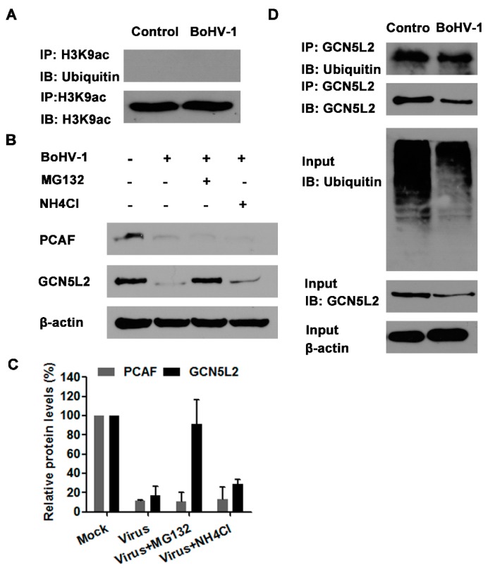
Figure 6.
The ubiquitin-proteasome pathway mediated GCN5L2 degradation. (A,D) MDBK cells in 60 mm dishes were mock infected or infected with BoHV-1 (MOI = 1) for 24 h. The cell lysates were prepared for IP using the antibody of either against H3K9ac (A) or GCN5L2 (D). The IP samples were subjected to immunoblots using antibodies against ubiquitin, H3K9ac and GCN5L2. The expression of GCN5L2 and ubiquitinated proteins in the input cell lysates in panel D were detected as a control. Data shown are representative of three independent experiments. (B) MDBK cells in 60 mm dishes were infected with BoHV-1 (MOI = 1) and treated with MG132 (1 μM), or mock treated with DMSO control for 24 h. The cell lysates were prepared for Western blotting to detect the expression of PCAF and GCN5L2. Data shown are representative of three independent experiments. (C) The band intensity was analyzed with software image J. Each analysis was compared with that of uninfected control which was arbitrarily set as 100%. The error bars denote the variability between the three independent experiments. +: indicated compound or virus was present, −: indicated compound or virus was not present.

