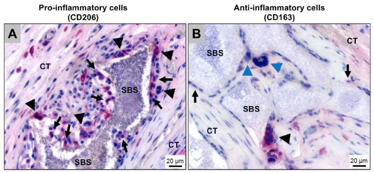
Figure 8.
Histological images showing the immunologic alignment of both macrophages and multinucleated giant cells (MNGCs) within the bony implantation bed of a synthetic bone substitute (SBS). CT: connective tissue. (A) Detection of pro-inflammatory mononucleated (arrows) and multinucleated cells (arrowheads) at the bone substitute granule surfaces (CD206-immunostaining); (B) Detection of anti-inflammatory mononuclear (arrows) and multinucleated (black arrowhead) cells at the bone substitute granule surfaces. Interestingly, most of the MNGCs that were adherent to the bone substitute granules (blue arrowheads) did not show an expression of the anti-inflammatory molecule (CD163-immunostaining).

