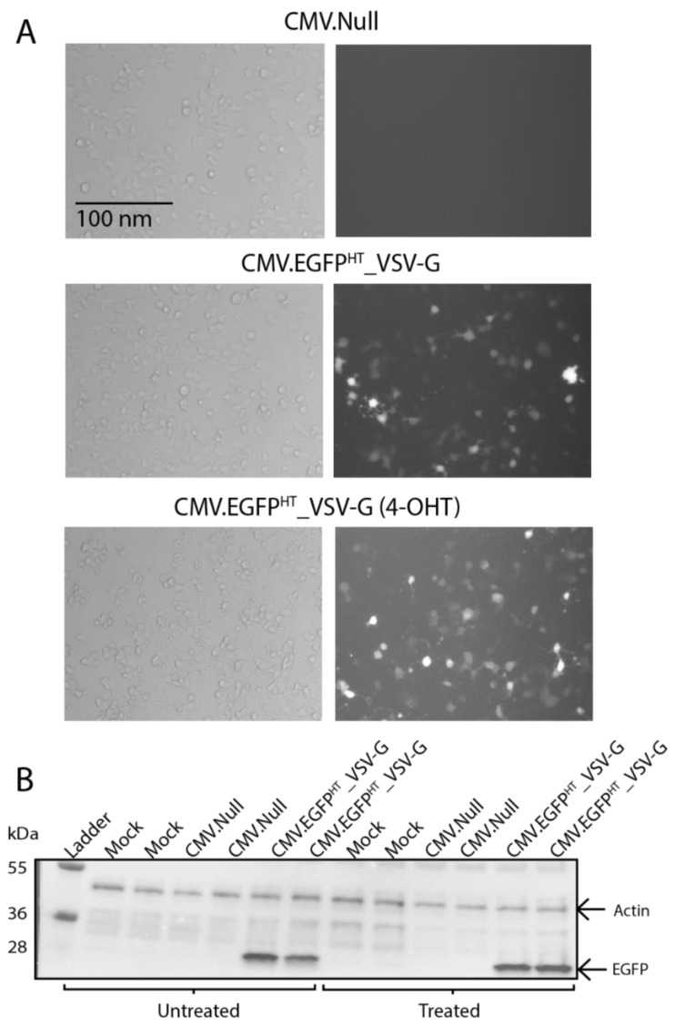
Figure 5.
(A) bright field (left) and fluorescence (right) images of tamoxifen-treated (4-OHT) and untreated EndoC-βH3 cells transduced with CMV.EGFPHT_VSV-G at MOI 250. Treated cells were incubated with tamoxifen for three weeks prior to transduction to remove the CRE-mediated immortalising transgenes. Images were taken at 48 hpt using a Zeiss Axiovert 135 inverted epifluorescence microscope (10×). Scale bar, 100 nm. (B) Tamoxifen-treated or untreated EndoC-βH3 cells were transduced with the indicated BacMam vectors or controls (in duplicate) and were harvested at 48 hpt for analysis by Western blotting using EGFP-specific antibody. β-actin was used as a loading control. Molecular weight markers are in kDa.

