Figure 1.
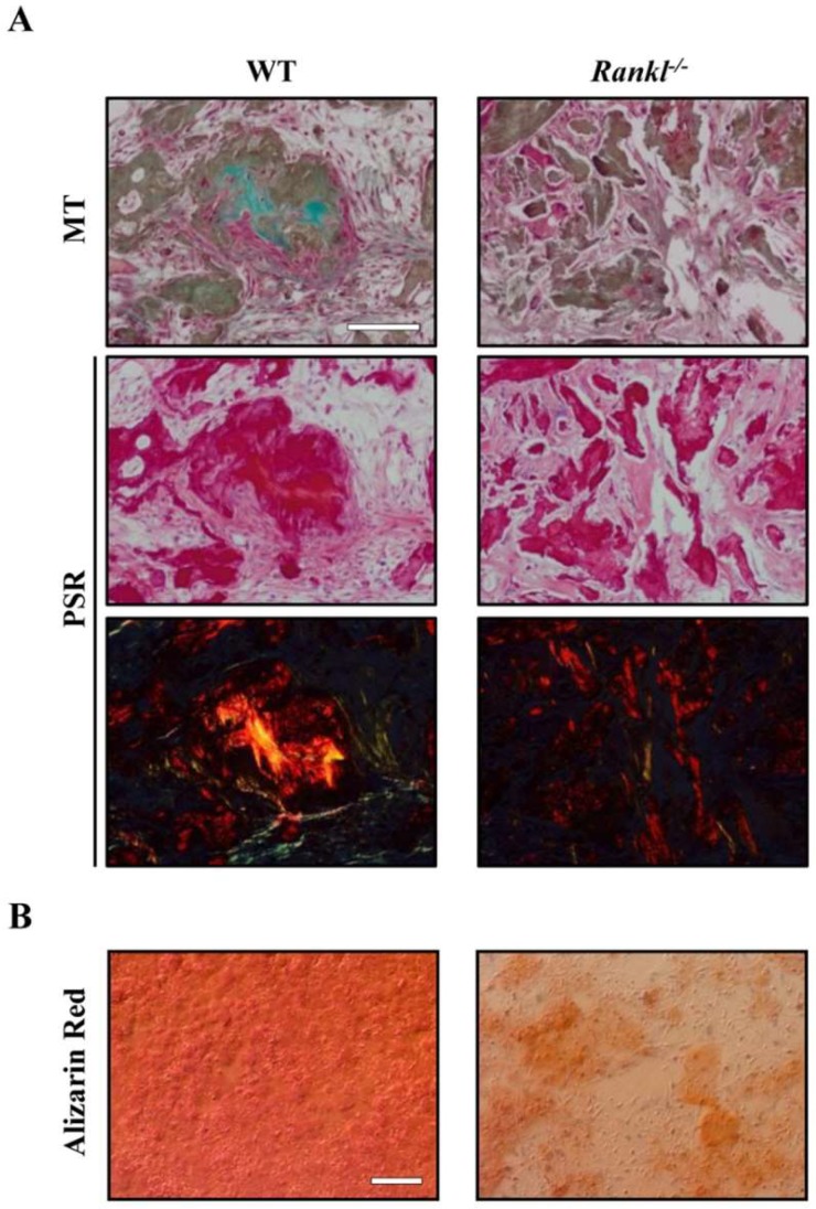
(A) Representative images of in vivo ceramic-based ectopic bone formation assay using wild type (WT) or Rankl−/− mesenchymal stem cells (MSCs). In scaffold systems seeded with WT MSCs, bone-like structures were present, as demonstrated by Masson’s trichrome (MT) staining of intense green collagen, and by yellow/orange birefringent fibers under polarized light in Picrosirius Red (PSR) staining. On the other hand, in Rankl−/− MSC-seeded scaffolds, the collagen deposition appeared less dense. Scale bar: 100 µm. (B) Representative images of in vitro WT or Rankl−/− MSC differentiation: MSCs were cultured in osteogenic medium for 14 days and mineralization was evaluated by Alizarin Red staining. Scale bar: 100 µm. Images are modified from [36].

