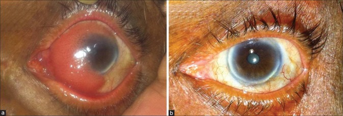
Figure 3.
(a) Preoperative clinical picture of tumor in Patient 3 with more than 3 quadrants of limbal area involvement, who underwent limbal stem cell transplantation in the form of conjunctivo-limbal autografting; (b) postoperative clinical picture showing stable ocular surface and no signs of limbal stem cell deficiency during follow-up

