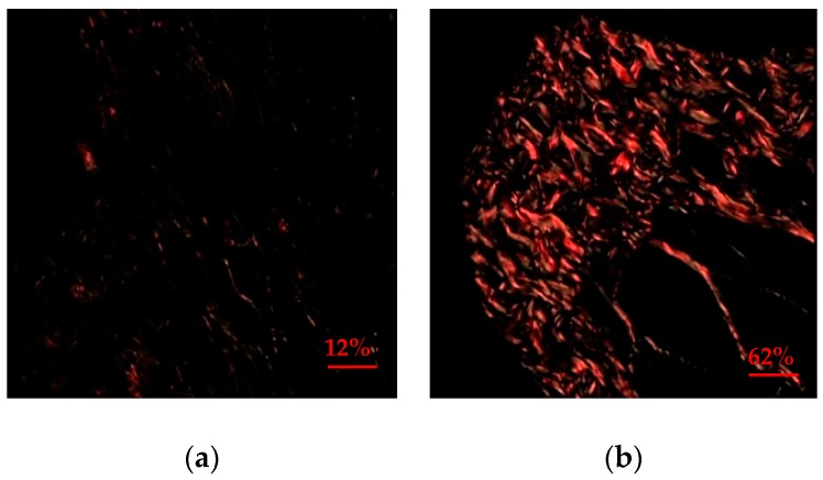
Figure 1.
Photomicrography of the expression of collagen in jugal mucosal of hamster obtained by picrossirius method under polarized light, in oral mucositis model at day 7, increase of 400×. (a) Photomicrography of the jugal mucosal, type III collagen, type 2, delicate and greenish fibers in the animals of the control group. (b) Photomicrography of the jugal mucosal, demonstrating type I collagen, thick and yellow-orange in animals of group supplemented with glycine.

