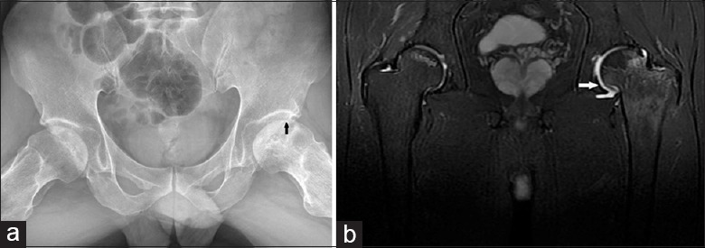
Figure 1.

Frog-leg lateral X-ray film shows a subchondral fracture (arrowheads) (a). Coronal T2-weighted fat-saturated fast spin-echo image (TR/TE: 1600/68) shows extensive bone marrow edema (arrowheads) in the adjacent right head and neck and joint effusion (b).
