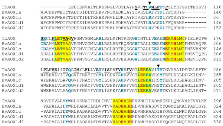
Figure 1.
Protein alignment of HvAOX isoforms with T. brucei AOX (TbAOX). Color scheme follows that previously established by Brew-Appiah et al. [12]: residues highlighted in yellow indicate conserved motifs. The residues bolded in red are amino acids proposed to coordinate the diiron center of the active site. Residues bolded in blue have been experimentally tested for loss of activity by previous researchers. Underlined and bolded residues are involved in the TbAOX hydrophobic cavity. The dark arrows indicate the residues R96 and T219.

