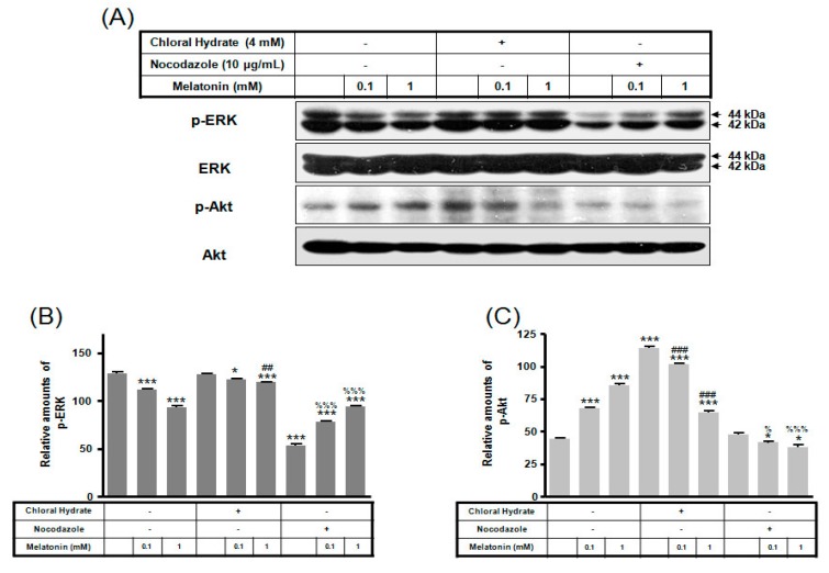
Figure 2.
p-ERK and p-Akt expressions in MC3T3-E1 cells with/without melatonin treatment. MC3T3-E1 cells were incubated in α-MEM supplemented with 10% FBS at 37 °C with 5% CO2. MC3T3-E1 cells in the presence or absence of melatonin (0.1, 1 mM) were treated with chloral hydrate (4 mM) for 3 days or nocodazole (10 μg/mL) for 4 h. p-ERK and p-Akt expressions were identified by Western blots (A). p-ERK (B) and p-Akt (C) expressions were quantified with ImageJ analysis software. * p < 0.05, *** p < 0.001 vs. FBS alone; ## p < 0.01, ### p < 0.001 vs. FBS + chloral hydrate; % p < 0.05, %%% p < 0.001 vs. FBS + nocodazole.

