Figure 1.
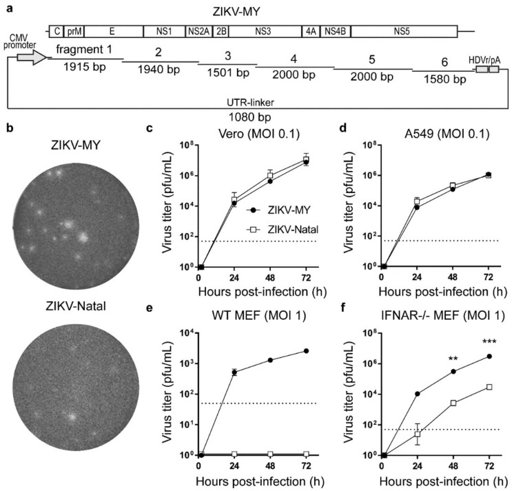
De novo generation and characterization of ZIKV-MY. (a) Schematic of Circular Polymerase Extension Reaction (CPER) fragments used for recovering ZIKV-MY. All fragments, except for the UTR-linker, are drawn to scale; (b) Plaque morphology on a Vero cell monolayer of ZIKV-MY recovered from the culture supernatant of CPER-transfected Vero cells, compared to ZIKV-Natal; Growth kinetics of ZIKV-MY versus ZIKV-Natal was performed on (c) Vero, (d) A549, (e) WT MEF, and (f) IFNAR−/− MEF cells at their indicated multiplicity of infection (MOI), and culture supernatants were harvested at the indicated time points post-infection and titered by plaque assay. The dashed lines represent the limit of detection of the assay. Means and ± SE are shown. Statistical analyses were performed using t-tests (n = three biological replicates); statistically significant are differences shown in panel (f) **—p = 0.008, ***—p < 0.001.

