Figure 7.
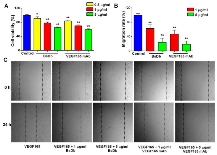
The recombinant BsDb inhibited VEGF-induced proliferation and migration of primary HUVECs. (A) BsDb inhibited the primary HUVECs proliferation. Primary HUVECs were cultured in 96-well plates and stimulated with 100 ng/mL VEGF165 and various concentrations of recombinant BsDb or VEGF165 mAb. Cell growth was then determined using an MTT assay; (B,C) BsDb inhibited the primary HUVECs migration. The HUVECs monolayer was scratched and placed in fresh Endothelial Cell Medium (ECM) containing 1% FBS with 100 ng/mL VEGF165 and various concentrations of recombinant BSDb or VEGF165 mAb. The HUVECs migration was photographed using a microscope after 24 h (40×) and the migration rate was calculated. The experiment was done in triplicate and the mean values ± SD were presented. * p < 0.05 and ** p < 0.01 using a two-tailed students t-test versus the control group.

