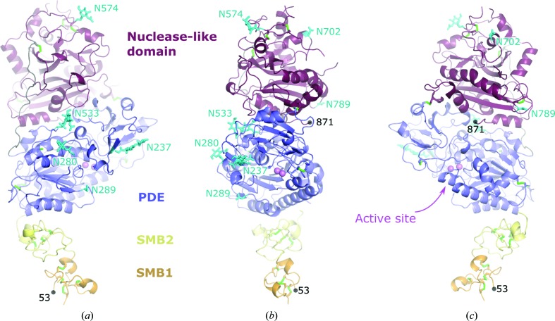
Figure 3.
Model of the full-length ectodomain of NPP3. (b) View from the front side, from which substrates have access to the active site (marked by the two zinc ions shown in magenta). Additional views are (a) from the left side [relative to the view in (b)] and (c) from the right side. This model was constructed by replacement of the PDE and nuclease-like domains of the NPP349–875 construct investigated in this work with the crystallographic coordinates of NPP3140–875 consisting of only the PDE and nuclease-like domains. Disulfide bridges are depicted in green and glycosylation sites in cyan. The N-terminal (53) and C-terminal (871) residues are marked.

