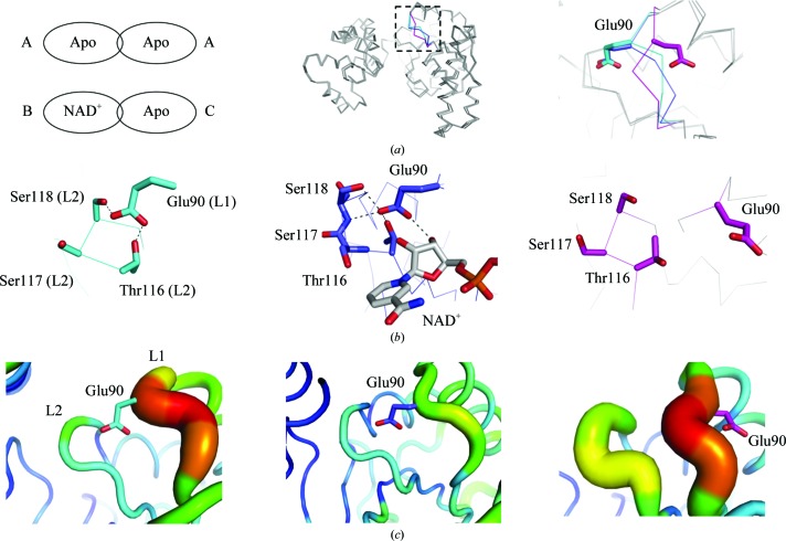
Figure 2.
Comparison of the monomer subunits in CacHBD. (a) Two types of dimers were found in the CacHBD crystal structures. They are schematically represented and the subunits are denoted as types A, B and C (left). 12 monomers in the asymmetric units of apo CacHBD and NAD+-bound CacHBD are superposed on the C-terminal domains. In the loop 1 region consisting of Ala87–Arg91, types A, B and C are partly colored cyan, blue and magenta, respectively (middle). Part of the dotted square in the middle panel is enlarged in the right panel. (b) Residues interacting with Glu90 are represented as stick models. The left, middle and right panels correspond to subunit types A, B and C, respectively. Dotted lines in the left and middle panels represent hydrogen bonds. (c) The B factor is shown in color, as prepared by PyMOL (Schrödinger). The highest and lowest values are shown in red and blue, respectively, with a gradient of colors in between. Each panel is viewed from the same direction as that in (b).

