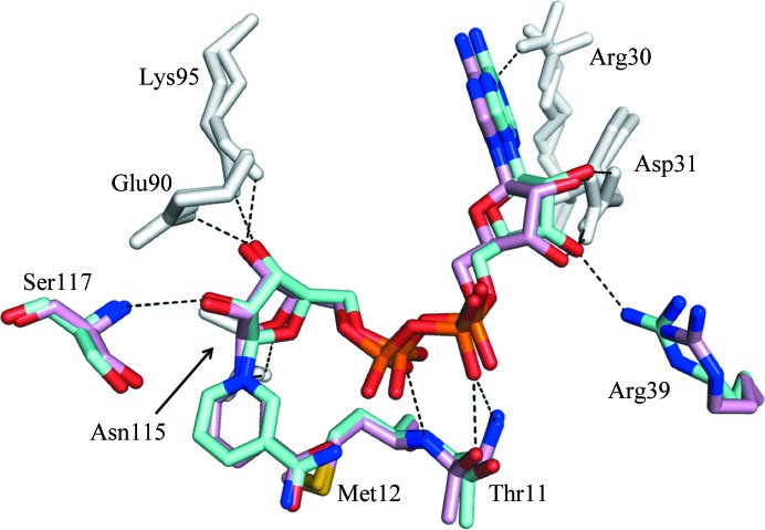
Figure 4.
Superposition of the NAD+-binding sites of CacHBD and CbuHBD. One of the two subunits of the NAD+-bound form of CacHBD (this study) and one of the four subunits of CbuHBD (PDB entry 4kug; Kim, Kim et al., 2014 ▸) are superposed and are colored cyan and pink, respectively, using Chimera. Residues colored gray represent the positions where hydrogen bonds are formed to NAD+ in all six subunits (two from CacHBD and four from CbuHBD). Residues colored cyan or pink indicate that the formation of hydrogen bonds depends on the subunits of both HBDs, with the exception that Arg39 formed a hydrogen bond to NAD+ only in CacHBD. Hydrogen bonds are represented by black dotted lines.

