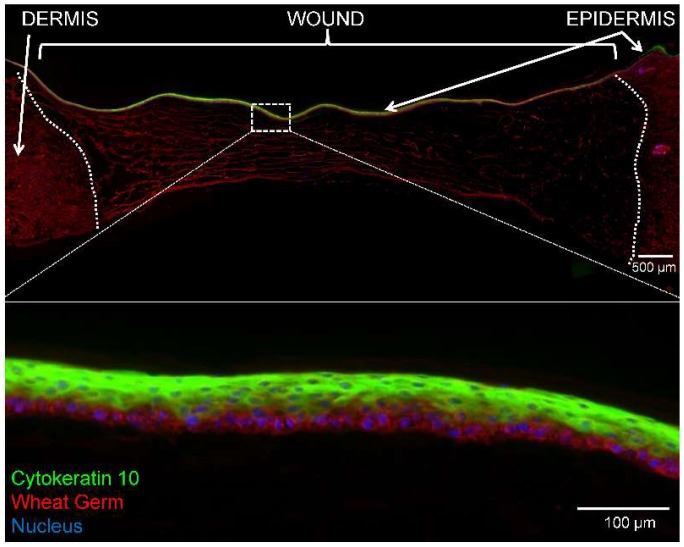
Figure 5.
Immunofluorescence of epidermal stratification on day 14 explants treated with PEG-PFP. (Top) CK-10 (green) stained the suprabasal layer of cells indicating formation of a stratified epidermis. Sections were counterstained with WG (red) to show the entire newly formed epidermis Nuclei (blue) are observed throughout the PEG-PFP hydrogel-treated area. The white dotted line represents the boundary from the normal dermis and the wound treated with PEG-PFP. (Bottom) Enlarged view of white boxed region with a better visual representation of the stratified epidermis from the center of the wound.

