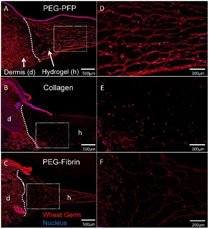
Figure 6.
Cellular migration into hydrogels. (A–C) Explants treated with different hydrogels were cryopreserved after 14 days in culture, sectioned, and stained with wheat germ (WG, red) to visualize all plasma membrane surfaces and counterstained with DAPI for nuclei (blue). Cells originating in the dermis of the explant can be seen infiltrating and migrating into the hydrogels. White dotted line represents the boundary of the normal dermal tissue (d) and hydrogel (h) in the wound. (D–F) Respective enlarged images of sectioned hydrogels demonstrate cellular density of migrating cells.

