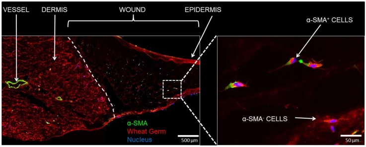
Figure 7.
Immunofluorescence of cellular infiltration. (Left) day 14 sections treated with PEG-PFP were stained with α-smooth muscle actin (α-SMA, green), a blood vessel and mesenchymal stem cell/myofibroblast marker, and counterstained with WG (red). The white dotted line represents the boundary between normal dermis and the wound treated with PEG-PFP. (Right) Enlarged view of white boxed region that shows cells from the normal tissue have infiltrated and proliferated into the hydrogel. Both α-SMA positive and negative stained cells were observed.

