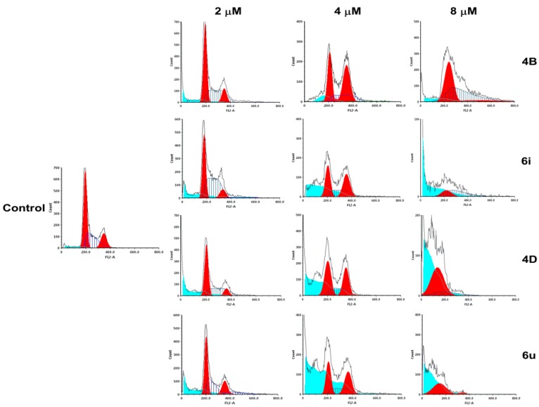
Figure 2.
The cell cycle was detected by flow cytometry using PI single staining. A549 cells were treated with 4B, 6i, 4D, and 6u at 2, 4, and 8 μM for 48 h, and then stained with PI for 30 min at 37 °C. The proportions of cells containing different DNA content were analyzed by FCS Express 5.0 (De Novo Software, Thornhill, Canada).

